Lamotrigine And Bipolar Depression.

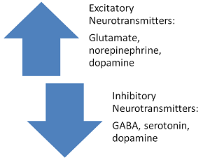
Lamotrigine is an anticonvulsant approved by the US Food and Drug Administration for maintenance mood stabilization of bipolar I disorder. Its acute antidepressant properties are recognized increasingly, both through meta-analysis and placebo-controlled evaluation. The anticonvulsant activity of lamotrigine is thought to be mediated by voltage-dependent sodium channel blockade, with subsequent presynaptic inhibition of aspartate (Asp) and glutamate (Glu) resulting in an overall reduction in neuronal excitability. Similar to lithium, lamotrigine also has been shown to be neuroprotective against Glu excitotoxicity in a number of cellular and animal models.
N-Acetylaspartate (NAA) is an abundant neuronal metabolite broadly, conceptualized as a marker of mitochondrial activity and neuronal integrity. Putative functions include: maintenance of brain fluid balance as an osmolyte, providing a source of acetate for myelin synthesis; serving as a precursor for synthesis of N-acetylaspartylglutamate (NAAG); and regulating Glu metabolism. NAA is synthesized in neuronal mitochondria from L-aspartate and acetyl coenzyme A through Asp N-acetyltransferase. An oligodendrocytic enzyme, aspartoacylase hydrolyzes NAA and thereby provides acetate for myelin synthesis. Glu and NAA metabolism are linked through the Glu-glutamine (Gln) and tricarboxylic acid cycles because such NAA may also serve as a Glu pool and buffer of glutamatergic excitoxcity.
The baseline deficit in NAA corrected with lamotrigine treatment is consistent with preclinical and animal models of lamotrigine-associated neuroprotection and NAA deficit normalization with lithium. For example, the NAA deficit, as a state marker, contributes to excessive glutamatergic tone and thereby leads to cell death and resultant decreases in NAA. The normalization of an NAA deficit could be mediated through activating Asp N-acetyltransferase, which promotes NAA synthesis. Also, lamotrigine may inhibit aspartoacylase, a catalytic enzyme in oligodendrocytes, or NAAG synthase in astrocytes. This increase in NAA levels may be associated with a reduction in Glu levels.
Postmortem brain studies consistently show reduced glial cell density in bipolar disorder. Rodent models of depression also show that compromised glial cell function induces depressive-like behaviors. Although NAA is synthesized in neurons and sensitive to mitochondrial integrity where it is synthesized, it is taken up and metabolized in glial cells. Therefore, reduced NAA levels in bipolar depression could relate in part to dysregulations in glial cell functioning or integrity, and this could be amenable to pharmacologic treatments.
In conclusion, after 12 weeks of lamotrigine treatment, NAA and tNAA levels normalized in patients with bipolar depression. This lamotrigine-associated brain change (i.e., increase in neuronal viability) may be related to increasing intraneuronal storage of Asp and, subsequently, NAA. Future research is encouraged to evaluate whether baseline NAA could be a potential biomarker for identifying lamotrigine response patterns and whether this functional brain change has an associated clinical response.