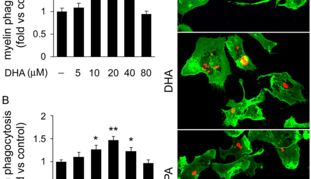
DHA or EPA.

BENEFICIAL EFFECTS OF DOCOSAHEXAENOIC ACID ON THE BRAIN.
Docosahexaenoic acid (DHA) is an important component of neural membranes and is present in 30-40% of the phospholipids in the gray matter of cerebral cortex and photoreceptor cells in the retina. It mediates its molecular and cellular effects not only through regulation of physicochemical properties such as membrane fluidity, permeability and viscosity in synaptic membranes, but also via modulation of neurotransmission, gene expression, and activities of enzymes, receptors and ion channels.
PUFAs including DHA have anti-oxidative stress, anti-inflammation and anti-apoptosis effects leading to neuroprotection in aged, damaged or Alzheimer’s disease brain.
Anti-oxidant and anti-inflammatory actions of DHA are associated with reduction in cellular levels of reactive species, pro-inflammatory mediators and nitrite levels, maintaining higher GSH levels and increasing anti-oxidant enzyme activities.
DHA attenuates brain necrosis after hypoxic ischemic injury by modulating membrane biophysical properties and maintaining integrity in functions between presynaptic and postsynaptic areas, resulting in better stabilization of intracellular ions after hypoxic-ischemic insult.
DHA also has a neuroprotective effect on glutamate-induced cytotoxicity in rat hippocampal cultures by inhibiting nitric oxide production and calcium influx, and increasing the activities of anti-oxidant enzymes glutathione peroxidase (GSH-Px) and glutathione reductase.
Many studies connect dietary consumption of DHA with antidepression. Areas with high consumption of seafood which is enriched in PUFAs such as DHA, have lower rates of bipolar and unipolar depression, post-partum depression and seasonal affective disorder compared to those where people consume less seafood.
DHA cannot be synthesized by the human body and must be taken in the diet. Common sources of n-3 PUFA include fish oils and some plant oils such as flaxseed oil and algal oil, which contain precursors of DHA, namely alpha-linolenic acid (α-LNA).
EFFECTS OF DHA ON NEUROTRANSMITTERS.
Glutamate.
DHA enhances glutamatergic synaptic activities with concomitant increases in synapsin and glutamate receptor subunit expression in hippocampal neurons. Spontaneous synaptic activity is significantly increased due to enhanced glutamatergic activity in DHA-supplemented neurons. On the other hand, lack of DHA results in inhibition of synaptogenesis, decreases in synapsins and glutamate receptor subunits, and impairment of long-term potentiation in hippocampal neurons. DHA modulates the activities of glutamate transporters (GLT1, GLAST, and EAAC1). DHA stimulates GLT1 and EAAC1 through a mechanism that requires extracellular Ca2+, CaM kinase II, and protein kinase C but not protein kinase A. In contrast, the inhibitory effect of DHA on GLAST does not require extracellular Ca2+ and does not involve CaM kinase II. DHA may also reduce uptake of the neurotransmitter d-[3H] aspartate by cultured astrocytes and cortical membrane suspensions, while AA (arachidonic acid) does not. This effect is also found in astrocytes enriched with the anti-oxidant, α-tocopherol, suggesting that it is not due to oxidation products of DHA. Reduction of d-[3H] aspartate uptake by DHA does not involve any change in the expression of membrane-associated astroglial glutamate transporters (GLAST and GLT-1), indicating that DHA reduces the activities of the transporters. In contrast to inhibition induced by free-DHA, membrane-bound DHA has no effect on d-[3H] aspartate uptake.
GABA.
DHA inhibits GABA receptor-mediated responses in cultured neural cells in a concentration and time-dependent manner.
Dopamine.
Chronic n-3 PUFA deficiency alters the internalization of dopamine (DA) in the storage pool of the frontal cortex. Diet-induced deficits in brain DHA lead to redistribution of DA vesicles in presynaptic terminals, greater basal extracellular DA concentrations and deficits in tyramine-induced elevations in extracellular DA concentrations in the prefrontal cortex and ventral striatum. Moreover, mice on a DHA deficient diet show augmented amphetamine-induced locomotor sensitization, and this response is associated with alterations in the mesolimbic DA pathway. These observations suggest that alterations in synaptic membrane DHA have an impact on DA synaptic neurotransmission and plasticity.
Noradrenaline.
Both a high membrane DHA content and free DHA in the medium enhance the release of [3H]-noradrenaline in cultured SH-SY5Y cells.
Acetylcholine.
Plasma [3H] DHA incorporation into synaptic membrane phospholipids of the rat brain is selectively increased after cholinergic activation.
DHA supplementation elevates brain DHA content, normalizes levels of brain-derived neurotrophic factor (BDNF), synapsin I, cAMP responsive element-binding protein (CREB), and CaMKII and improves learning ability in rats after traumatic brain injury. DHA deficit induces down-regulation of brain glucose transporter protein expression, resulting in decreased glucose utilization in the cerebral cortex of DHA-deficient rats. DHA also interacts with the peroxisome proliferator activated receptors (PPAR), sterol regulatory element binding protein (SREBP), and carbohydrate response element binding protein (ChREBP). These influence the activities of signal transduction processes during neurotransmission.
Benefits of EPA.
The ultimate goal of using omega-3 fatty acids is the reduction of cellular inflammation. Since eicosanoids derived from arachidonic acid (AA), an omega-6 fatty acid, are the primary mediators of cellular inflammation, EPA becomes the most important of the omega-3 fatty acids to reduce cellular inflammation for a number of reasons. First, EPA is an inhibitor of the enzyme delta-5-desaturase (D5D) that produces AA (1). The more EPA you have in the diet, the less AA you produce. This essentially chokes off the supply of AA necessary for the production of pro-inflammatory eicosanoids (prostaglandins, thromboxanes, leukotrienes, etc.). DHA is not an inhibitor of this enzyme because it can’t fit into the active catalytic site of the enzyme due to its larger spatial size. As an additional insurance policy, EPA also competes with AA for the enzyme phospholipase A2 necessary to release AA from the membrane phospholipids (where it is stored). Inhibition of this enzyme is the mechanism of action used by corticosteroids.
Finally, it is often assumed since there are not high levels of EPA in the brain, that it is not important for neurological function. Actually it is key for reducing neuro-inflammation by competing against AA for access to the same enzymes needed to produce inflammatory eicosanoids. However, once EPA enters into the brain it is rapidly oxidized (2,3). This is not the case with DHA (4). The only way to control cellular inflammation in the brain is to maintain high levels of EPA in the blood. This is why all the work on depression, ADHD, brain trauma, etc. have demonstrated EPA to be superior to DHA.
Summary.
EPA and DHA do different things, so you need them both, especially for the brain. If your goal is reducing cellular inflammation, then you probably need more EPA than DHA. How much more? Probably twice the levels, nonetheless you always cover your bets with omega-3 fatty acids by using both EPA and DHA at the same time. Both seem to be equally effective in making powerful anti-inflammatory eicosanoids known as resolvins.